
Industry-academia collaboration promoted research in qualitative analysis of myocardial blood flow and evaluation of ischemia using CT and MRI and resulted in software for analyzing myocardial blood flow. It clearly explains the relation between coronary artery stenosis and myocardial ischemia (Figure 1).
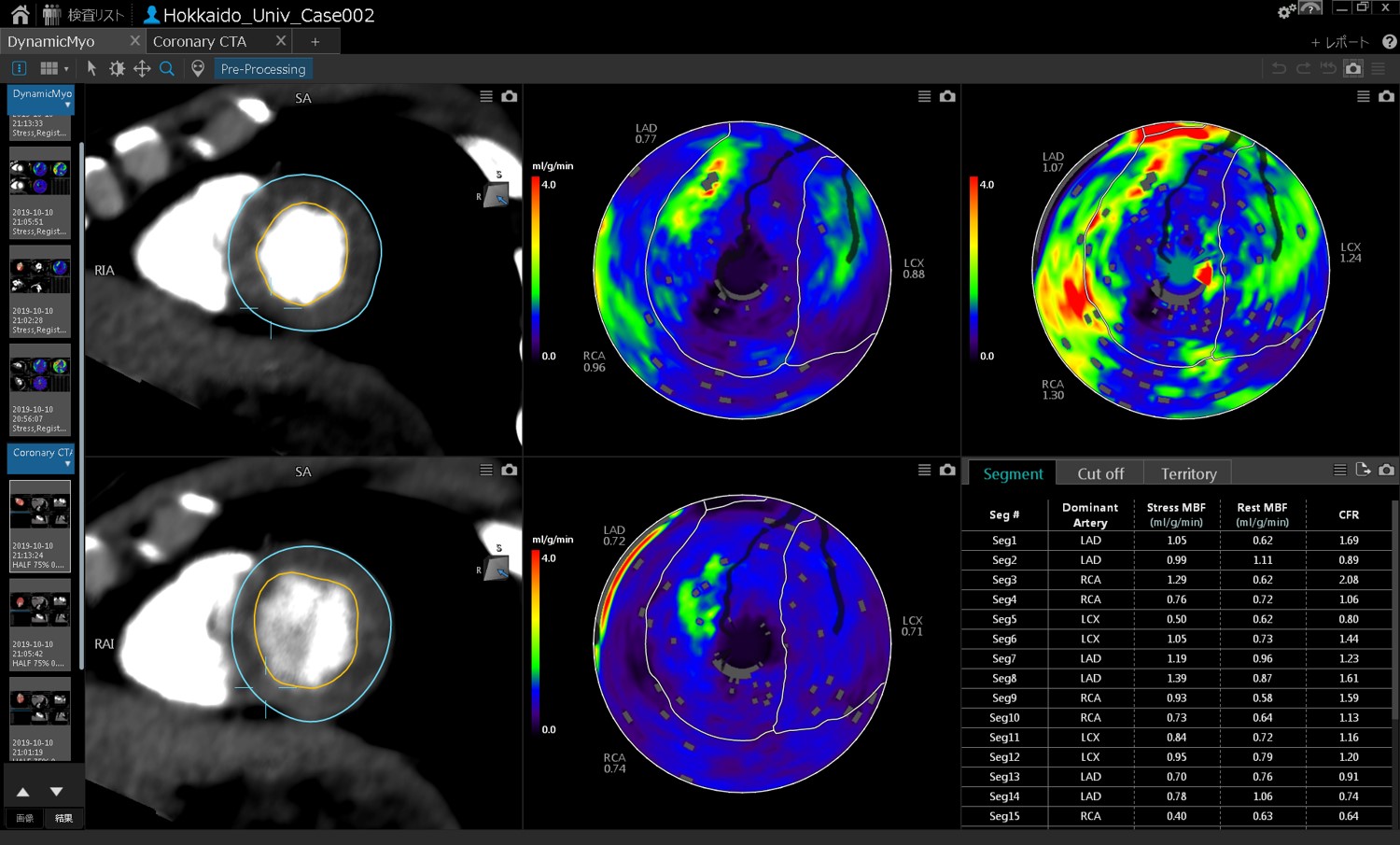
This image represents ischemia in the right coronary artery and anterior descending branch of left coronary artery. A perfusion range is indicated for each artery depending on the artery size. It is easier to detect which artery is responsible for ischemia.
4D-flow MRI visualizes blood flow without using a contrast agent and without exposure to radiation. This imaging technique is being examined for quantitative evaluation of valvular heart disease and vascular malformation.
We succeeded in visualizing blood flow kinetics of complex vascular malformation for the first time in the world. Our research appeared on the cover of a leading international scientific journal.
We hold a regular joint conference with the Department of Cardiovascular Medicine on radiologic interpretation of cardiac MRI and coronary CT to learn quantitative evaluation and the basics of radiologic interpretation.